- Dr. Mattioli
- Image analysis
- Specialisations
- Surgical procedures
- Face
- Surgery
Rinoplasty

Blepharoplasty

Otoplasty

Mentoplasty
THE FORMS OF BEAUTY
THE FACE IS THE WINDOW TO THE SOUL
(MARCUS TULLIUS CICERO, DE ORATORE, 55 BC)

Home/Face/ears
The outer ear consists of the pinna, commonly referred to as ear, which collects sound vibrations and guides them into the auditory canal towards the thin membrane of the eardrum.
The ear pavilion is composed of cartilage finished with a thin layer of skin. The external aspect is an irregular oval, meaning that it does not avail of a flat surface since the cartilaginous blade acts as a support frame to the protruding parts and cavities.
The helix is the outermost of the protrusions and is composed of a sheaf that follows the margin of the pavilion towards its external face which ends up in the lobe at the rear, and before that arises from the root of the helix.
The antihelix is a concentric cartilaginous sheaf that goes towards the helix and arises from two branches that unite to form the trunk and delimit a cavity called the triangular cavity.
On the upper part of the helix there may be a thickening either inwardly or outwardly, called Darwin’s tubercle.
The ear’s protrusion is defined in terms of distance and angle. In the normal ear the angle of separation from the cranium is at about 30°, with a mean distance at the upper edge of the helix from the cranium surface of about 1.5 – 2 cm.
The prominent ear originates from an embryonal alteration, in failing to form the fold of the antihelix.
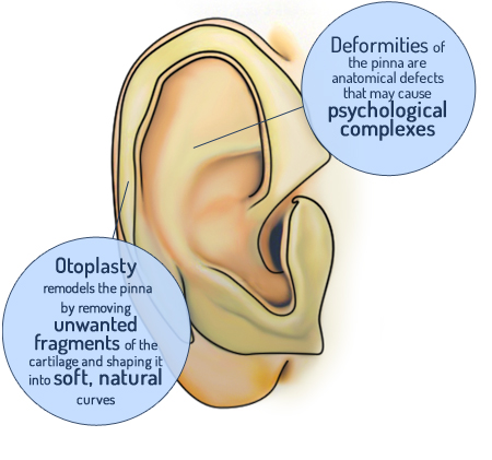
The malformation of the ear pavilion whether due to hypertrophy of the basin or failure to fold the antihelix (these alterations may in some cases appear together) is an anatomical defect that may determine psychological complexes.
The surgical techniques usually used for otoplasty procedures, entail the remodeling of the ear cartilage through the removal of fragments from the basin or the creation of gentle and natural curves with suture points in the convex zones.
Ecco la moderna chirurgia del naso con la funzionalità e l'estetica
Clinica Sant'Anna, interview with Dr. Mattioli

I thought it was going to be much more painful